Hey there, fellow biotech folks and lab gurus! Let’s cut to the chase—you’re running a host cell residual protein (HCP) ELISA or whatever detection kit you’re using, and suddenly the results are all wonky. Maybe your positive control is too high, your samples are coming up with weird spikes, or your standard curve looks like a lopsided rollercoaster instead of a smooth line. I get it, I’m on the kit supply end of this game, and I’ve heard every HCP troubleshooting horror story from labs just like yours. Today, I’m walking you through the stuff that actually fixes this garbage—no fancy jargon, no dead ends, just real steps that work when your HCP results are off. Host Cell Residual Protein Detection Kits

First off, let’s ground this: HCP kits (whether ELISA, Western blot, or Luminex) are super sensitive, which is why they’re such workhorses for biomanufacturing—you’re looking for trace leftover proteins from E. coli, CHO, whatever your host is, and even tiny mess-ups in protocol or materials will blow up your data. I’ve seen new lab techs panic because their sample looks dirty, only to find they contaminated their own standards. Let’s start with the low-hanging fruit that almost every lab misses first.
Wait, hold up—before you even touch your samples or reagents, check your kit storage. This is so basic it’s almost embarrassing how many labs mess this up. Most HCP antibodies and detection reagents are temperature-sensitive, right? I’ve had customers send me emails like “I left my kit on the bench overnight because I got swamped” and wonder why their control signal is half what it should be. Most of our kits need 2–8°C for storage, and some of the lyophilized components (like standards or conjugates) need to be frozen long-term—def don’t leave those out in a warm lab for hours. Pro tip: if you’re not using all components at once, split any thawed reagents into single-use aliquots and stick them back in the fridge. Thawing and refreezing over and over breaks down antibodies and standards, which totally skews your calibration curve.
Next up, your sample prep. This is the #1 culprit for weird HCP results, I swear. HCPs are everywhere—your buffers, your pipette tips, your labware, even your gloves. Wait, did you use regular pipette tips that aren’t filter tips? A lot of labs skip filter tips to save money, but they’re cheap compared to contaminated samples. I’ve had a customer come to me with CHO HCP results that were 10x higher than expected, and it turned out their lysis buffer had residual protein from a bad batch of buffer they bought off Amazon (don’t do that, just use our recommended diluents, okay? We test our diluents to be HCP-free). Also, are you diluting your samples correctly? If you under-dilute, you can get matrix effects—stuff in your final sample (like cell culture media components) will block antibody binding, making your HCP read falsely low. Over-dilute, and your HCP is below the kit’s limit of detection, so your standard curve can’t pick it up. Rule of thumb: do a quick pre-test dilution (1:100, 1:500, 1:1000) to nail the sweet spot where your samples fall within the standard curve range. Oh, and if your samples are really viscous? Maybe you need to filter them through a 0.22μm filter—viscosity can make them stick to the plate or pipette tips, leading to inconsistent volumes. That’s another big one I see all the time.
Now, let’s get to the actual assay setup—this is where I see so many small mistakes add up. First, pipetting errors. I’ve lost count of labs that use the wrong pipette for small volumes (like using a P1000 for 5μL transfers) and get uneven liquid across the plate. Even if you have a fancy pipette, you need to calibrate it every 3–6 months—cheap, easy, and avoids half your wells having too much or too little reagent. Also, did you mix your reagents properly? Conjugates and standards settle at the bottom of their tubes, so you need to invert them gently (don’t vortex like a maniac, that can break antibodies) for 30 seconds before using. If you skip mixing, your standards will have uneven protein concentrations, leading to a wonky curve. Another thing: incubation times and temperatures. Our HCP ELISA kits, for example, need a 1-hour incubation at 37°C, not 45 minutes at room temp, and definitely not 2 hours on the bench while you grab coffee. Even a 15-minute mismatch in incubation time can change signal by 20–30%, which is enough to make your results unusable. Also, make sure your plate reader is set to the right wavelength—for most of our kits, that’s 450nm, but some have a reference at 620nm to correct for plate background. If you forget to set the reference, you might get extra noise from dirty wells that look like real signal.
Wait, what about the controls? If your positive control (the one with known HCP concentration) is giving weird results, that’s a way easier fix than your samples, so start there. If your positive control is way too high: did you accidentally use the standard instead? No, just kidding, but seriously—check labels, controls vs. standards are different. Also, if you’re reusing controls? Don’t, that’s a bad call. If the control is too low: it’s probably degraded, which goes back to the storage thing I mentioned earlier. Or maybe you forgot to add the antibody conjugate to the control wells—super common, I’ve done it myself once during a 16-hour assay run. Another control red flag: high background in the blank well (the one with no HCP, just diluent). That means non-specific binding—either your well plate is dirty (make sure you’re not touching the bottom/inner edges of the plate with your gloved hands, grease from skin causes background), or your wash step was bad. Oh, wash steps! People skip this all the time. For ELISA, you need to wash 3–5 times, each time leaving the wash buffer for 30 seconds before dumping it, and slapping the plate dry on lint-free paper (don’t rub, that damages the wells). If you don’t wash enough, unbound conjugate stays in the well, leading to random high signals everywhere. That’s such a simple fix, but it’s the first thing I check when a customer says their blanks are spiking.
Now, the trickier stuff: matrix effects. If your samples are in a complex matrix (like cell culture supernatant, fermentation broth, or purified protein drug substance), the stuff around your HCP can mess with the antibody’s ability to bind. I’ve seen this so much with mAb purification—your purified mAb is fine, but the feed stream has media components that block the antibodies in the kit, making HCP read falsely low. How do you fix this? Use matrix-matched standards! Instead of diluting your HCP standards in our kit’s generic diluent, dilute them in your sample matrix (i.e., the same cell culture media or buffer you use for your samples). That accounts for the matrix’s effect, and gives you accurate numbers. We actually have tips for this in our kit manuals, so if you haven’t read the manual cover-to-cover yet—stop scrolling, go read it. I know manuals are boring, but they have all the stuff for troubleshooting, plus we test every kit with common matrices so we know what works.
Wait, what if your standard curve is flat or doesn’t fit? That’s usually either pipetting error when making the serial dilutions of standards, or the standards are degraded. When you make serial dilutions, always change pipette tips between each dilution—cross-contaminating standards will make your curve look weird. Also, don’t make your standards too far in advance—they’re only stable for a few hours once thawed and diluted, so make them right before you run the assay. If all else fails, reach out to the kit supplier (hi, that’s me!)—we run the same tests in our quality control lab every day, so we can tell you if the kit is faulty or if it’s a protocol mistake. I can’t tell you how many times a customer sends a weird curve, I ask a few quick questions (storage, prep, incubation time) and it’s a simple thing they missed. For example, last week a biotech lab had a curve that only had two points on it—turns out they used a 96-well plate that had leftover reagent from a previous assay, so half the wells didn’t work. Easy fix, once we asked.
Also, let’s talk about common pitfalls specific to different kit types. If you’re using a Western blot HCP kit, your transfer might be bad—protein didn’t transfer from gel to membrane, so your bands are faint or missing. If you’re using a Luminex kit, you might have microparticle aggregation, so your counts are all over the place. For Luminex, make sure you sonicate the microparticles gently before use, and don’t let them sit at the bottom of the tube without mixing. Again, all these little steps are in the kit insert—we don’t bury this stuff on purpose, we write it there for a reason.
Wait, one more big one: contamination from previous experiments. If you’re running HCP assays multiple times a week, your bench, pipettes, or incubator might have leftover HCP from old samples. That’s called carryover, and it’s a huge issue for sensitive kits. How do you fix this? Decontaminate your bench with 70% ethanol before every assay, use dedicated pipettes for HCP work (no using the same pipette for cell culture and HCP samples), and if you have a big carryover issue, use a 10% bleach solution to wipe down your incubator or plate shaker—bleach breaks down proteins, so it gets rid of leftover HCP. I had a customer who was getting 50ng/mL HCP in their blank wells, and it turned out their incubator had a bottle of E. coli lysate spilled on the shelf a week earlier. Bleach cleaned it, and their blanks went back to normal.
Let’s wrap this up with a quick checklist you can tape to your bench next time you run an HCP assay:
- Check all reagent storage (no leaving stuff out!)
- Prep samples with filter tips, dilute to the right range, filter viscous samples
- Calibrate pipettes, mix reagents gently, change tips for standards
- Follow incubation times/temps exactly, don’t skip wash steps
- Run matrix-matched standards, use fresh controls
- Decontaminate your bench before starting
And if you run through all this and still get weird results? Don’t stress—reach out to the kit supply team. We’re here to help, not just sell you boxes of kits. We can send you a replacement kit if ours is faulty (we rarely have issues, but mistakes happen) or walk you through step-by-step troubleshooting over a quick call. At the end of the day, accurate HCP results are non-negotiable for biomanufacturing, vaccine development, or any protein work—so don’t let a small mistake sink your project.
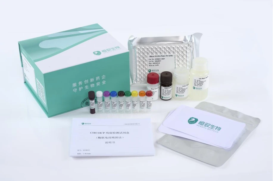
If you’re gearing up for your next HCP test, or you’re tired of dealing with wonky results that delay your work, let’s chat about your needs. We can help you pick the right kit for your host cell type, send over a full troubleshooting guide tailored to your work, and get you set up with what you need to get reliable data fast. Stop wasting time on messed-up assays—let’s make this simple.
Host Cell Residual Protein Detection Kits References
- Tomanicek, S.J., et al. (2019). Troubleshooting host cell protein detection in biopharmaceutical manufacturing. Journal of Pharmaceutical and Biomedical Analysis, 171, 112678.
- Rosenberg, A.S. (2005). Immunogenicity of protein therapeutics: the role of host cell proteins. Nature Biotechnology, 23(8), 1007–1013.
- Liu, Y., et al. (2021). Critical factors affecting accuracy of ELISA-based host cell residual protein quantification. Analytical Biochemistry, 623, 114157.
HyQure Biotech Co., Ltd.
As a professional host cell residual protein detection kits enterprise in China and Switzerland, we rely on international compliance standards, leverages excellent quality and technical strength. If you have any enquiry about cooperation, please feel free to email us.
Address: Building 18, Lane 2777 East Jinxiu Road, Pudong New Area, Shanghai 2.Stücki Park Hochbergerstr. 60 I, 4057 Basel Switzerland
E-mail: service@hyqure.com
WebSite: https://www.hyqure.com/
